Protein Structure — free PNG images to download
Ribbon diagrams of real protein folds: coiled alpha helices, short beta strands and loose loops, most of them coloured along the chain from blue at one end to red at the other, a few with stick-model side groups.
- Images
- 7
- Sizes
- 800 to 1 200 px
- Formats
- PNG with alpha, WebP, JPG
- Download
- free, no sign-up
Filters
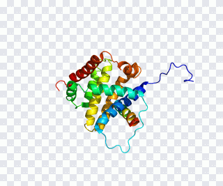 BCL2L11 Bcl-2 family BCL2-like 1 Protein PNG
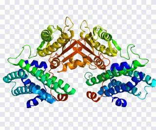 PDK4 Pyruvate dehydrogenase kinase Pyruvate PNG
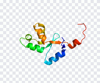 OCA2 hect domain and RLD 2 Gene Protein PNG
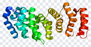 FANCE FANCA Fanconi anemia, complementation PNG
 Melanocortin 4 receptor Melanocortin PNG
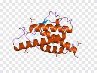 Interleukin 9 Interleukin 4 Crystal PNG
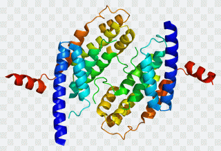 TERF2 Telomere Protein TERF1 Encyclopedia PNG
Reading the colours
The rainbow shading is not decoration. In molecular graphics it traces the chain from its start (blue) to its end (red), so a viewer can follow how the sequence folds back on itself. Single-colour or two-tone models usually separate the subunits of a complex instead.
Monomers, dimers and receptors
Some images show a compact single domain, others a symmetrical pair of chains or a long receptor threading through a membrane in a row of helices. Pick the shape that matches your subject: a dimer for enzyme kinetics, a membrane bundle for signalling and pharmacology, a small globular fold for an introduction to protein structure.
Using them in teaching
The ribbons sit on a transparent background, so they can be laid over a cell diagram or placed beside a sequence alignment without a white box around them. They read well at slide size; for posters, choose the files with fewer, thicker helices.
Questions and answers
Is the transparency real or a painted checkerboard?
All 7 images carry a real alpha channel. On the image page you can switch the backdrop to white or dark to see the edges.
Are there large files for print?
The largest files in this set reach 1 200 px. For print choose 2000 px and up in PNG; for the web 500–800 px in WebP.
Do I need to register to download?
No sign-up needed. The file is served after a short wait on the download page.
Can I use protein Structure images commercially?
The catalogue gathers images from open sources. Before commercial use confirm the rights with the author — the source link is on the image page.