Enzyme — free PNG images to download
Ribbon models of enzymes: rainbow-shaded folds, two-chain dimers in red and blue, sky-blue beta sheets ringed by orange helices, and sprawling multi-domain complexes.
- Images
- 6
- Sizes
- 800 to 1 200 px
- Formats
- PNG with alpha, WebP, JPG
- Download
- free, no sign-up
Filters
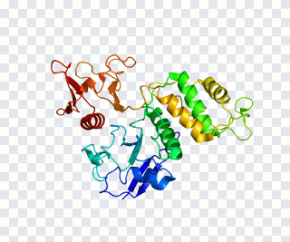 PTK2B Integrin-linked kinase Gene Protein PNG
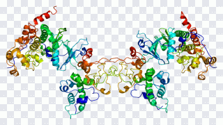 Casein Kinase 2 Alpha 1 Text, Csnk2b PNG
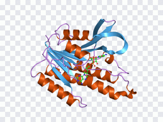 Short-chain dehydrogenase Reductase Enzyme PNG
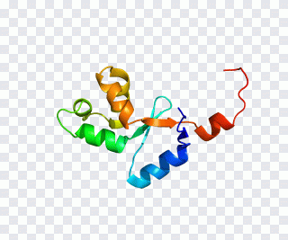 OCA2 hect domain and RLD 2 Gene Protein PNG
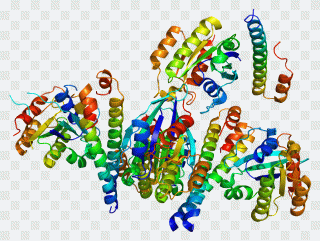 P-glycoprotein Gene Rabaptin RABEP1, text PNG
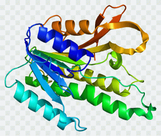 QDPR Phenylalanine hydroxylase Gene PNG
Reading a ribbon
Coils are alpha helices, flat arrows are beta strands, and thin threads are the loops between them. The rainbow shading runs along the chain from one end, usually blue, to the other, usually red, so it shows where the protein starts and how it folds back on itself.
Picking a model
A compact single fold is the clearest choice for a textbook figure or a quiz about secondary structure. A symmetric dimer explains how identical subunits pair up, and the large multi-coloured complexes give a sense of scale for slides on cellular machinery.
On a page
These renders come with transparent backgrounds, so they sit well over a pale gradient or a lab photo. On a dark cover the deep blue segments fade, and a light backing keeps the whole chain visible.
Questions and answers
Are all enzyme images really transparent?
Yes. Every file is checked for an alpha channel rather than a white or chequered backdrop. Images without real transparency do not enter the catalogue.
What sizes are available?
The largest files in this set reach 1 200 px. For print choose 2000 px and up in PNG; for the web 500–800 px in WebP.
How many files can I download?
Downloads are free and open. With very frequent requests the site asks to confirm you are not a robot — a normal visitor never sees it.
What is the licence?
Terms depend on the specific image. We neither sell files nor issue licences; for commercial work check the source named on the image page.